
Who is it for?
Kidney ablation is an alternative to surgery, particularly for small renal tumors (masses staged T1a, generally under 4 cm in international guidelines). It is especially valuable for patients with a single kidney, limited renal function, high surgical risk due to comorbidities, or who decline surgery.
Suitability is assessed from tumor size, location and overall health, and decided together with the urology team. In appropriate cases a biopsy documents the tumor type before or during the session.
How it is done — step by step
Planning
The tumor is mapped with ultrasound and CT as needed; a safe distance from neighbouring organs is planned (fluid can be instilled to widen the gap when needed).
Anaesthesia
Performed under sedation or with the anaesthesia team for comfort.
Ablation
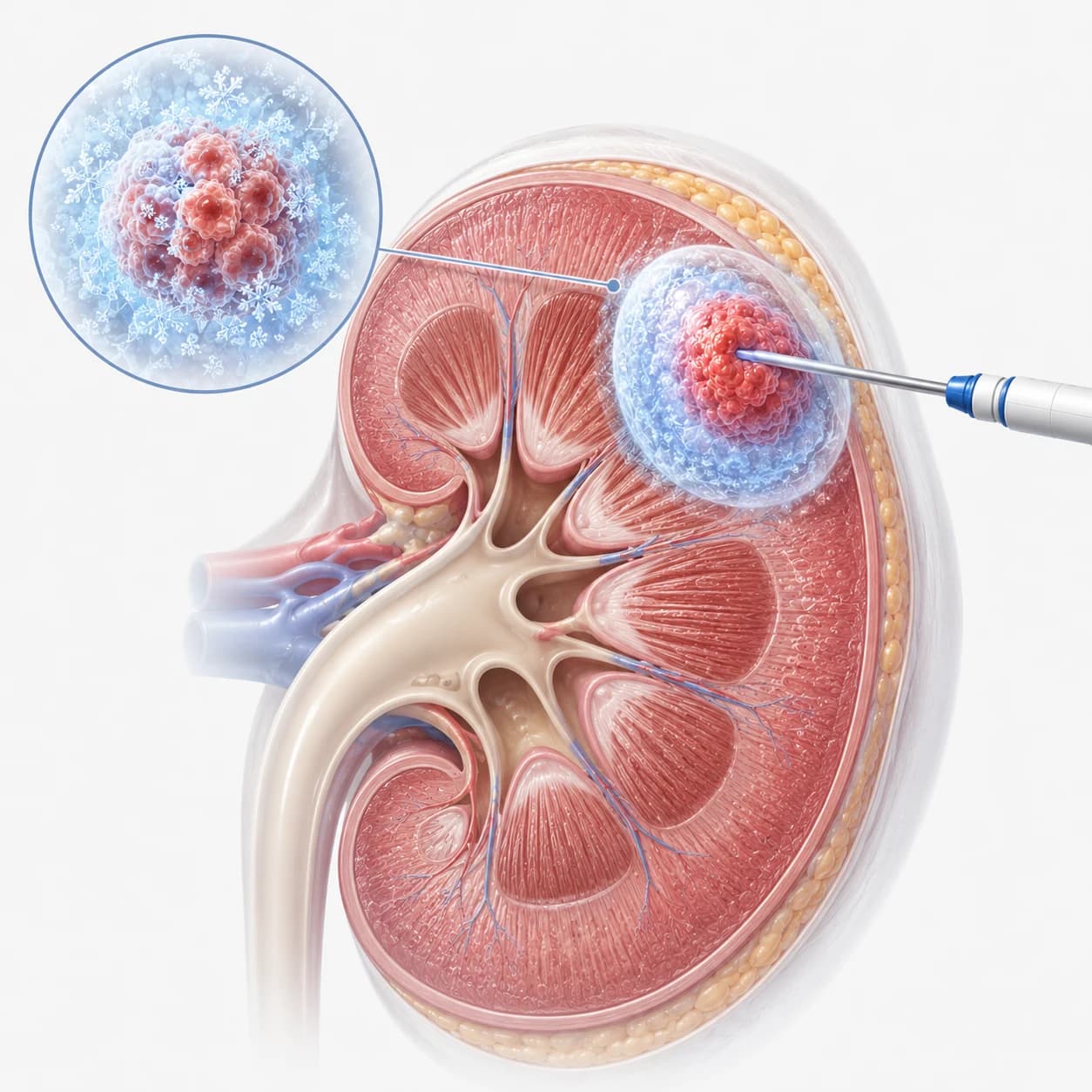
Under image guidance a needle-antenna is placed in the tumor; microwave/radiofrequency heat (or freezing in selected cases) inactivates the tumor with a safety margin.
Check
The ablation zone is imaged at the end; only a needle mark remains on the skin.
What to expect on the day
You will be asked to fast; blood thinners are adjusted in advance. The procedure usually takes 30–90 minutes.
You are observed for several hours; some patients go home the same day, others after one night. Transient pink urine can occur.
Risks and Limitations
Flank soreness and fatigue may last a few days. Bleeding is rare; injury to the urinary tract or neighbouring organs is very rare and minimized with planning and protective techniques.
Limits: success decreases for large or centrally located tumors, where surgery may be preferable. Recurrence can occur after ablation — regular imaging follow-up is an integral part of the treatment, and the procedure can be repeated if needed.
Recovery and follow-up
Most patients return to daily life within days; avoiding heavy lifting for the first week is sufficient.
Follow-up uses CT or MRI at set intervals to monitor both the treated zone and the rest of the kidney; the schedule is built with the urology team.
Scientific References
- AUA — Küçük böbrek kitleleri kılavuzu (Campbell SC ve ark., J Urol, 2021)
- EAU — Böbrek hücreli karsinom kılavuzu (Ljungberg B ve ark., Eur Urol)
- Dernek kaynakları: CIRSE (Avrupa Girişimsel Radyoloji Derneği) · SIR (ABD) · SIO (Society of Interventional Oncology) · TGRD (Türk Girişimsel Radyoloji Derneği)
Frequently Asked Questions
Will my kidney be harmed?
A key advantage of ablation is preservation of healthy kidney tissue; function loss is typically less than with surgery — critical for patients with a single kidney.
Which tumors are suitable?
Primarily small (generally under 4 cm), favourably located tumors. Suitability is assessed on imaging together with urology.
How does it differ from surgery?
No incision or organ removal, faster recovery, and repeatability. Conversely, surgery may be the better option for large or central tumors.
Will I feel pain during the procedure?
No — it is done under sedation or anaesthesia care. Post-procedure flank soreness is well controlled with medication.
What if it recurs?
If follow-up imaging shows an active focus, ablation can usually be repeated; surgery is reconsidered when needed.
Will a biopsy be taken too?
In appropriate cases, yes — before or during the same session, to document the tumor type and plan long-term follow-up correctly.
The information on this page is for general education; diagnosis and treatment decisions are made individually after clinical evaluation.