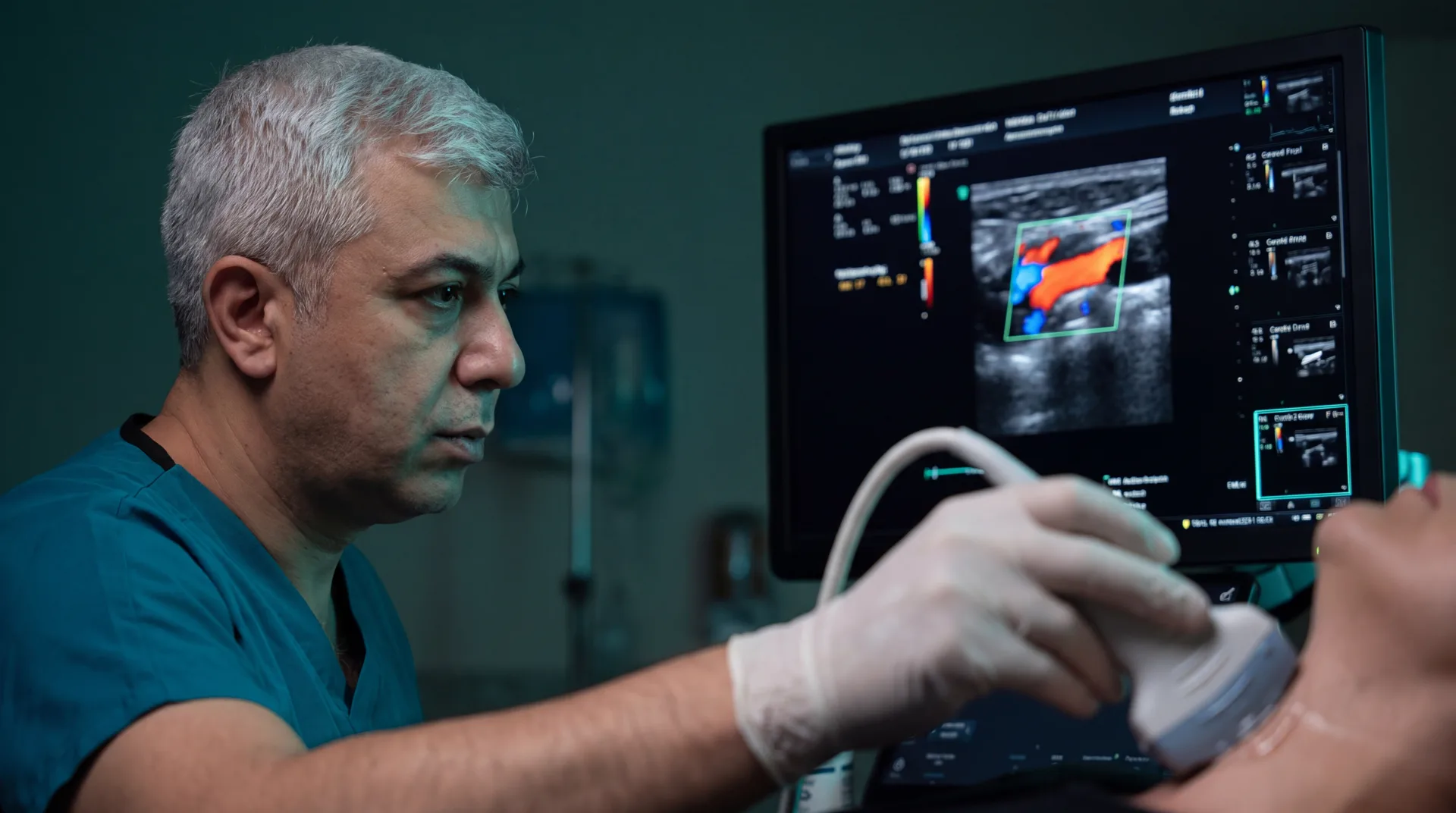
How Is It Performed?
Ultrasonography uses high-frequency sound waves — without any radiation — to image internal organs in real time. Doppler ultrasound additionally measures the direction and velocity of blood flow, enabling assessment of vascular disease and organ perfusion.
Advantages
- No radiation — safely repeatable
- Painless and non-invasive
- Real-time, dynamic assessment
- Expert-level interpretation and reporting
- Initial findings shared in the same session
What Does the Process Look Like?
Preparation
Depending on the region examined, simple preparations such as short fasting or a full bladder may be requested; you will be informed when booking.
Examination
The area is scanned with a probe after gel is applied. The examination usually takes 15–30 minutes and is completely painless.
Reporting
Findings are shared with you after the examination and a detailed written report is prepared.
Frequently Asked Questions
Is ultrasound harmful?
Ultrasonography involves no radiation; it can be used safely and repeatedly in all age groups, including pregnant women and children.
How should I prepare for the examination?
Abdominal ultrasound usually requires a few hours of fasting; pelvic examinations may require a full bladder. Thyroid and superficial tissue examinations need no preparation.
What does a Doppler examination show?
Doppler evaluates vascular conditions such as stenosis, occlusion, varicose veins and organ blood supply.
When will I get the results?
Preliminary findings are usually shared at the end of the examination; the detailed written report is prepared shortly afterwards.
The information on this page is for general education; diagnosis and treatment decisions are made individually after clinical evaluation.