
Who is it for?
Lymph node biopsy identifies the cause of enlarged or suspicious nodes in the neck, armpit, groin or abdomen: infection, benign reactive enlargement, lymphoma or spread from another tumor can only be distinguished by tissue examination.
The method follows the suspicion: fine-needle aspiration for cytology, core needle when tissue architecture matters (particularly suspected lymphoma); some cases need surgical excision. The choice is made with the relevant specialties.
How it is done — step by step
Assessment
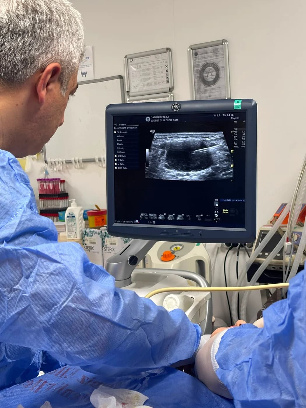
The target node is examined with ultrasound; the most suspicious, safest sampling point is chosen.
Local anaesthesia
Skin and track are numbed.
Sampling
Samples are taken with a fine or core needle under ultrasound guidance; each pass lasts seconds. Extra material is set aside for special tests (such as flow cytometry) when needed.
Finish
Brief compression and a small dressing; no stitches.
What to expect on the day
Fasting is usually unnecessary; tell us in advance if you take blood thinners.
The visit typically takes 15–30 minutes; after brief observation you return to your day.
Risks and Limitations
Superficial node biopsy is low-risk: mild soreness and bruising are most common; significant bleeding and infection are rare. In vessel-rich regions such as the neck, continuous ultrasound vision protects these structures.
A limit: needle biopsy may not allow definitive classification of some lymphoma types; surgical removal of the whole node may then be required. This possibility is discussed from the start.
Recovery and follow-up
Normal life resumes the same day; site tenderness settles within days.
Results, including any special studies, are usually complete within days to two weeks and are reviewed with your treating physician.
Scientific References
- SIR — Perkütan iğne biyopsisi kalite iyileştirme kılavuzu (Gupta S ve ark., J Vasc Interv Radiol, 2010)
- ATA — Erişkinlerde diferansiye tiroid kanseri yönetim kılavuzu (Ringel MD, Sosa JA ve ark., Thyroid, 2025)
Frequently Asked Questions
Is every enlarged node bad?
No — nodes also enlarge with infections, and most enlargements are benign. Biopsy brings clarity when suspicious features persist.
Fine needle or core?
Cytology may need only a fine needle; core is preferred when architecture matters, as in suspected lymphoma. The choice follows the suspicion and the node’s location.
Is it painful?
Done under local anaesthesia; most patients feel only pressure, and it is over in minutes.
When is the result ready?
Usually within days to two weeks — special studies for lymphoma can extend this slightly.
Will surgical biopsy be needed?
Definitive classification of some lymphomas requires excision of the whole node; needle biopsy is the first and often sufficient step.
Can I return to work the same day?
Yes — after brief observation you can resume your day.
The information on this page is for general education; diagnosis and treatment decisions are made individually after clinical evaluation.